

Contents
hide
Congenital cataracts mean your baby is born with a cloudy area in the lens of their eye. This cloudiness can impair clear vision during the first weeks of life, a very important time for your baby’s eyesight to develop. If these cataracts aren’t treated within the first 4 to 10 weeks, your child could have permanent vision loss. That’s why it’s so important to find them early, get surgery if needed, and keep up with regular eye check-ups.
What Are Congenital Cataracts?
Congenital cataracts are a clouding of the lens that develops before or just after birth. Unlike cataracts in adults, which usually develop slowly over time, congenital cataracts can affect a baby’s vision right away, during the first weeks and months of life.
A baby’s brain learns to see during the first weeks and months after birth. If a cataract causes blurred or blocked images, the connection between the eye and the brain may not develop properly. This is why early treatment is so important; delaying surgery can result in permanent vision problems.
Why Congenital Cataracts Matter

Even a small cataract can affect a baby’s vision. Babies need clear images to learn to focus, follow movement, and see depth.
This may lead to:
- amblyopia (lazy eye)
- poor depth perception
- Reduced vision in one or both eyes
- long-term developmental impact if untreated
A small cataract in the center of the lens can change how a baby’s vision develops.
Why Cataracts Develop Before Birth
The lens of the eye forms early in pregnancy. Problems with genes, infections, or metabolism at this time can cause the lens to become cloudy. Causes include:
1. Genetic Factors
Many congenital cataracts are inherited. Some affect only the eyes, while others are part of genetic syndromes involving other organs.
Conditions that may include cataracts:
- Down syndrome
- Marfan syndrome
- Weill–Marchesani syndrome
- Stickler syndrome
- Lowe syndrome
- Myotonic dystrophy
Most congenital cataracts are inherited. If one parent has the condition, it can be passed to their child.
2. Infections During Pregnancy
Some infections can pass from mother to baby during pregnancy and affect lens development. These TORCH infections include:
- toxoplasmosis
- syphilis
- rubella
- cytomegalovirus (CMV)
- herpes simplex
Rubella infection during early pregnancy is still a common cause of congenital cataracts.
3. Metabolic or Systemic Disorders
Metabolic or systemic disorders can cause cataracts soon after birth. Examples include:
- galactosemia (a metabolic emergency)
- hypocalcemia
- Amino-acid metabolism disorders
- rare forms of neonatal diabetes
Finding these problems early is important for protecting both vision and overall health.
How Congenital Cataracts Appear
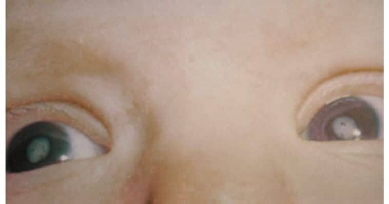
Parents are often the first to notice signs of a problem. These signs include:
- a white or dull reflection in the pupil (leukocoria)
- lack of eye contact or poor tracking
- abnormal eye movements (nystagmus)
- one eye turning inward or outward
- a baby who doesn’t visually explore their surroundings
In many cases, the first sign is a white pupil in a photograph, especially with flash photography.
If you notice any of these signs, a pediatric eye doctor should check your child as soon as possible.
How Eye Doctors Diagnose Congenital Cataracts
Because babies cannot describe what they see, pediatric ophthalmologists rely on specific tests to understand how well the eye is developing.
- Check the red reflex (the small reddish glow you see when a light is shined into the pupil)
- Look closely at the lens using magnification to see how clear it is
- determine whether light is reaching the retina normally
- Examine the rest of the eye to make sure all structures are healthy.
If the cataract is dense and the posterior segment cannot be visualized, ocular ultrasound is used to assess the retina and other intraocular structures.
Depending on your child’s needs, the doctor may order more tests:
- blood tests to check for infections or metabolic conditions
- genetic testing if an inherited cause is suspected
- ERG or VEP testing to assess how the retina and optic nerve react to light
The eye doctor will ask about your pregnancy, your child’s birth, and if anyone in your family has had eye problems. This information helps the doctor find out if the cataract is the only issue or part of a bigger health problem. It also helps plan the best care for your child.
Most of these tests are painless and can be performed during a routine eye exam.
Types of Congenital Cataracts
Congenital cataracts can develop in different areas of the lens. The type and location of the cataract determine how much vision is affected and how quickly treatment may be needed. Types of congenital cataracts include:
Total Cataract
A total cataract means the whole lens is cloudy. Very little light can reach the retina, so vision is affected from birth. Surgery is usually needed as soon as possible to help vision develop normally.
Nuclear Cataract
Nuclear cataracts affect the central part of the lens, called the nucleus, which helps focus light. Cloudiness in this area causes blurred vision. These cataracts are often inherited and may occur in several family members.
Lamellar (Zonular) Cataract
Lamellar cataracts form in layers or rings around the lens’s center. Some are thin and cause little vision loss, while others are thicker and more noticeable. Vision may be normal or blurry, depending on the density of the cataract. These cataracts often develop during a specific stage of pregnancy and may remain stable or slowly worsen over time.
Cerulean Cataract
Cerulean cataracts appear as small blue-white dots in the lens. They are often harmless and usually do not change over time. These cataracts are often found during routine eye exams. Most children with cerulean cataracts have good vision and do not need surgery.
Posterior Polar Cataract
Posterior polar cataracts appear as a round, dense white spot at the back of the lens. Even a small spot in this area can cause vision problems. Surgery for this type of cataract is more delicate because the cloudy area is attached to thin structures behind the lens.
Anterior Polar Cataract
Anterior polar cataracts form at the front of the lens as a small, clear spot. They usually cause little or no vision problems and can be monitored with regular eye exams. Most children with this type do not need surgery and develop normal vision with ongoing check-ups.
Why Identifying the Type Matters
Knowing the type of congenital cataract helps the doctor predict:
- How much vision might be affected?
- whether amblyopia (lazy eye) may develop
- how quickly surgery or treatment is needed
- What visual rehabilitation (glasses, patching) will be required
A thorough eye exam and early check-up are important for planning the best treatment for your child’s vision.
Treatment: When Surgery Is Needed
Small cataracts that do not block the center of vision can often be monitored with regular eye exams. Dense or central cataracts usually require early surgery to protect your child’s vision.
Typical timing guidelines:
- One eye affected: ideally by 4–6 weeks of age.
- Both eyes are affected: typically by 8–10 weeks.
- Partial cataracts: Surgery is only needed if vision worsens over time.
How Infant Cataract Surgery Works
Cataract surgery in babies is different from surgery in adults because a baby’s eye:
- heals faster
- responds more strongly to inflammation
- grows significantly after surgery
The procedure may include:
- a tiny incision in the cornea or limbus
- removal of the cloudy lens
- opening the back of the capsule
- a small anterior vitrectomy to prevent clouding
- placement of a weaker intraocular lens (IOL) or no lens at all
Some babies use contact lenses after surgery until they are old enough to get a permanent lens implant.
Timing is important. Early surgery gives the brain clear images and helps normal vision develop.
After Surgery: Vision Development Begins
Removing the cataract is the first step. After surgery, the brain needs to learn to use the eye to see.
Post-surgery care may include:
- anti-inflammatory and antibiotic drops
- regular pressure checks
- glasses or a special infant contact lens
- amblyopia therapy (patching the stronger eye)
Vision therapy and regular follow-up visits help vision develop.
Many parents find the time after surgery challenging. With regular follow-up and support, most children adapt well.
Possible Complications
Even after successful surgery, children with congenital cataracts can have long-term risks, such as:
- glaucoma (can develop months or years later)
- recurrent clouding of the visual axis
- inflammation or membrane formation
- IOL displacement
- rare retinal detachment
Complications can happen months or years after surgery. Regular eye exams are needed for life.
These complications are not caused by anything parents did. They occur because infant eyes heal differently and continue to grow after surgery.
Outlook for Children With Congenital Cataracts
With early detection and appropriate treatment, many children with congenital cataracts can achieve good vision. Children with cataracts in both eyes often do better than those with cataracts in only one eye, because both eyes develop together.
Outcomes depend on:
- timing of surgery
- the type of cataract
- presence of the other eye or health conditions
- consistency with patching and follow-up care
Parents play an important role in their child’s vision. Taking part in treatment and follow-up visits is important.
Can Congenital Cataracts Be Prevented?
Not all congenital cataracts can be prevented, but there are some things you can do to lower the risk:
- Rubella vaccination before pregnancy
- good prenatal care
- screening for maternal infections
- red reflex testing for all newborns
- genetic counseling for at-risk families
Finding cataracts early is the best way to protect your child’s vision and give them the best chance for healthy sight.
FAQs
Can congenital cataracts come back after surgery?
The cataract itself doesn’t return, but sometimes a thin, cloudy layer can form behind the new lens or in the visual pathway. This is called visual axis opacification. If it happens, a simple and quick procedure can usually restore clear vision for your child.
Are congenital cataracts always easy for parents to notice?
Not always. Some cataracts are tiny or not in the center of the lens, so they’re hard to spot just by looking. That’s why doctors check every newborn’s eyes with a special light, called a red reflex test, to catch cataracts that parents might not see.
Can children live a normal life after treatment?
Absolutely. If a child receives early surgery and regular follow-up care, most can see, learn, and play just like other kids. Some children may need vision therapy for a few years, but with support, they can thrive in everyday life.
Can congenital cataracts be a sign of other health issues?
Sometimes, yes. Congenital cataracts can sometimes be linked to other health conditions, such as genetic, metabolic, or infectious problems. That’s why doctors may suggest extra tests, just to make sure your child is healthy in every way.
Is cataract surgery safe for babies?
Yes, when done by a specialist who treats children. The surgery is generally safe, but your child will need regular check-ups afterward, because some issues, like glaucoma, can develop as they grow. Ongoing care helps catch any problems early.
Summary
Congenital cataracts are a clouding of the lens in the eye that is present at birth or in early infancy. They can disrupt normal visual development and may lead to permanent vision loss if left untreated.
Early diagnosis, surgery within the first 4 to 10 weeks, and regular exams with glasses, contact lenses, and patching give your child the best chance for healthy vision.