Contents
hide
The optic nerve connects your eyes to your brain. If it’s harmed, you can lose vision. Most people don’t think about it until something feels wrong. Understanding the optic nerve helps you catch problems early.
Before we look closer, it’s important to understand the optic nerve’s essential role in your vision. This guide explains its function, how it works, the conditions that can affect it, symptoms to watch for, and steps to protect your vision.
What Is the Optic Nerve?

The optic nerve is part of your central nervous system (the system that controls your brain and spinal cord). It contains over one million tiny nerve fibers that transmit visual information from the retina, which is a layer of light-sensitive cells at the back of your eye, to your brain.
Each eye has its own optic nerve. Together, these nerves form the pathway that allows you to recognize faces, read text, perceive colors, and detect movement.
The optic nerve is surrounded by protective layers known as meninges. The meninges are the same coverings that protect your brain and spinal cord. Because of this close connection, certain conditions affecting the brain can also impact your vision.
Where the Optic Nerve Begins: The Optic Disc
The optic nerve starts at the optic disc, also called the optic nerve head. This is the location where the nerve fibers exit the eye. The optic disc creates a natural blind spot because it lacks light-sensing cells. The brain compensates for this, making people usually unaware of the gap.
During a dilated eye exam, your doctor will check your optic disc for swelling, paleness, or changes in shape. These can be signs of a problem.
How the Optic Nerve Works
The optic nerve carries messages from your eye to your brain. Each step in this process is important for a clear vision. Here is how the process works:
Light First Enters the Eye
Light enters your eye, passes through the clear front surface called the cornea, and then through the lens, before reaching the retina at the back of the eye.
The Retina Converts Light Into Electrical Signals
The retina contains two main types of cells: rods and cones. Rods help you see in low light and provide peripheral vision, while cones allow you to see colors and fine details.
These cells convert light into electrical signals that your brain can interpret.
Signals Collect and Enter the Optic Nerve
All of these signals collect at the optic disc, where they join together and travel down the optic nerve toward your brain.
The Optic Chiasm: Where Nerve Fibers Cross
Deep within your brain, the optic nerves from each eye meet at a structure called the optic chiasm.
At the optic chiasm, some of the nerve fibers cross over to the opposite side of the brain. This crossing is important for depth perception and for coordinating vision from both eyes.
At the optic chiasm, some of the nerve fibers cross over to the opposite side of the brain. This crossing is important for depth perception and for coordinating vision from both eyes.
Without this crossing of nerve fibers, you would not be able to perceive three-dimensional depth.
Signals Reach the Visual Cortex
After passing through the optic chiasm, the signals travel to the visual cortex located at the back of your brain. The visual cortex processes these signals and creates the images you see.
Anatomy of the Optic Nerve: A Closer Look
The optic nerve measures about 3.5 to 5 centimeters in length (about the size of your little finger). It is divided into four main segments, or parts:
Intraocular Segment (Inside the Eye)
This segment includes the optic disc (the spot where nerve fibers leave the eye) and the lamina cribrosa (a mesh-like structure that the nerve passes through). These areas are particularly vulnerable to damage from glaucoma.
Intraorbital Segment (Behind the Eye)
This flexible segment lies behind the eye and allows your eye to move in different directions without straining the optic nerve.
Intracanalicular Segment
This segment passes through the optic canal, a narrow space that can make the nerve more susceptible to compression.
Intracranial Segment (Inside the Brain)
This section runs inside the brain and connects the optic nerve to the optic chiasm, continuing the visual pathway that carries signals to your brain.
Various diseases can affect different segments of the optic nerve. The symptoms you experience will depend on which part of the nerve is involved.
Conditions That Can Affect the Optic Nerve
There are many potential causes of optic nerve damage. Early detection is important, as damage to the optic nerve is often permanent.
Glaucoma
Glaucoma damages the optic nerve, often due to increased pressure inside the eye. Vision loss usually starts at the edges of your visual field and can progress unnoticed.
Glaucoma damages the optic nerve, often due to increased pressure inside the eye. Vision loss usually starts at the edges of your visual field and can progress unnoticed.
Optic Neuritis
Optic neuritis is frequently associated with autoimmune diseases. It may cause pain when you move your eye, make colors appear less vivid, or create a dark spot in your vision.
Optic neuritis is frequently associated with autoimmune diseases. It may cause pain when you move your eye, make colors appear less vivid, or create a dark spot in your vision.
Optic Disc Drusen
Small calcium deposits in the optic nerve head, known as optic disc drusen, can cause the nerve to appear irregular and may affect your peripheral vision.
Small calcium deposits in the optic nerve head, known as optic disc drusen, can cause the nerve to appear irregular and may affect your peripheral vision.
Ischemic Optic Neuropathy
A sudden decrease in blood flow to the optic nerve is called ischemic optic neuropathy. This means the nerve does not get enough oxygen-rich blood, which can cause sudden, painless vision loss.
A sudden decrease in blood flow to the optic nerve is called ischemic optic neuropathy. This means the nerve does not get enough oxygen-rich blood, which can cause sudden, painless vision loss.
Compressive Optic Neuropathies
Tumors, cysts, thyroid disorders, or other growths can compress the optic nerve. This pressure can gradually worsen vision.
Tumors, cysts, thyroid disorders, or other growths can compress the optic nerve. This pressure can gradually worsen vision.
Medication-related or toxic injury
Certain medications and severe vitamin deficiencies can also damage the optic nerve.
Certain medications and severe vitamin deficiencies can also damage the optic nerve.
Symptoms of Optic Nerve Problems
If your optic nerve is damaged, vision may look dim or faded. Colors may seem dull, and objects less clear.
- You may notice blurry vision or difficulty reading small print as fine details become harder to see.
- If the optic nerve becomes swollen, you may experience eye pain.
- Loss of peripheral (side) vision can be an early sign of optic nerve problems.
- You may notice blank spots or shadows in your field of vision.
- It may become more difficult to see in low-light conditions, making activities such as night driving or walking in dark areas more challenging.
If you experience sudden vision loss, seek medical attention immediately.
If you develop sudden eye pain, nausea, halos around lights, or a rapid decrease in vision clarity, seek prompt medical care.
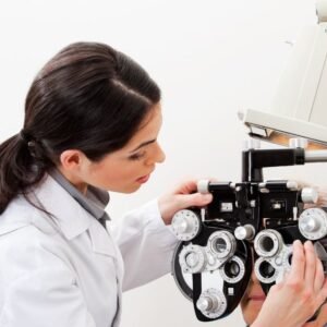
How Doctors Diagnose Optic Nerve Problems
A comprehensive eye examination can detect whether your optic nerve is swollen, compressed, or damaged.
• Complete eye examination
Your eye doctor will examine your optic nerve through your pupil to look for swelling, changes in shape, or paleness.
Your eye doctor will examine your optic nerve through your pupil to look for swelling, changes in shape, or paleness.
• Visual field testing
This test can reveal areas where your vision is missing or reduced. It is useful for detecting glaucoma and other optic nerve disorders.
This test can reveal areas where your vision is missing or reduced. It is useful for detecting glaucoma and other optic nerve disorders.
• Optical Coherence Tomography (OCT)
An OCT scan is a painless test. It measures the thickness of the nerve fiber layer in your eye. It can show if your optic nerve is swollen or thinner than normal.
An OCT scan is a painless test. It measures the thickness of the nerve fiber layer in your eye. It can show if your optic nerve is swollen or thinner than normal.
• MRI of the brain and orbits
An MRI can find swelling, tumors, high pressure, infection, or other problems near your optic nerve.
An MRI can find swelling, tumors, high pressure, infection, or other problems near your optic nerve.
• Blood testing
Blood tests can find autoimmune diseases, infections, or vitamin deficiencies that can affect the optic nerve.
Blood tests can find autoimmune diseases, infections, or vitamin deficiencies that can affect the optic nerve.
Treatment Options
Treatment for optic nerve problems depends on the cause. Common treatments are:
• Pressure-lowering therapy for glaucoma
Prescription eye drops, laser treatment, or surgery can lower eye pressure and help protect the optic nerve.
Prescription eye drops, laser treatment, or surgery can lower eye pressure and help protect the optic nerve.
• Anti-inflammatory treatment
If you have optic neuritis or another condition that causes swelling, your doctor may give you steroids to reduce inflammation and help the nerve recover.
If you have optic neuritis or another condition that causes swelling, your doctor may give you steroids to reduce inflammation and help the nerve recover.
• Managing systemic illness
Controlling your blood pressure, diabetes, infections, or autoimmune diseases can help prevent more damage to the optic nerve.
Controlling your blood pressure, diabetes, infections, or autoimmune diseases can help prevent more damage to the optic nerve.
• Removing or shrinking compressive masses
If you have a tumor or other growth, treatment may include surgery or radiation.
If you have a tumor or other growth, treatment may include surgery or radiation.
• Medication changes
If a medicine is affecting your optic nerve, your doctor may change or stop it.
If a medicine is affecting your optic nerve, your doctor may change or stop it.
• Vision rehabilitation
If you have permanent optic nerve damage, tools such as magnifiers or specialized vision training can help you stay safe and independent.
If you have permanent optic nerve damage, tools such as magnifiers or specialized vision training can help you stay safe and independent.
How to Protect Your Optic Nerve
Adopting healthy habits can help reduce your risk of optic nerve damage.
- Maintain good control of your blood pressure and diabetes.
- Avoid smoking.
- Include foods rich in B vitamins and omega-3 fatty acids in your diet.
- Wear protective eyewear to reduce the risk of eye injuries.
- Schedule comprehensive eye examinations every one to two years, or more frequently if you are at higher risk.
- Consult your doctor promptly if you experience headaches or other neurological symptoms.
- Begin regular glaucoma screenings after the age of 40.
When to See an Eye Doctor Immediately.
Certain symptoms require you to see an eye doctor right away for urgent evaluation.
- Sudden vision loss
- Severe eye pain
- A new blind spot
- Loss of color brightness
- Vision dimming with headache (possible papilledema)
- Visual distortions or flickering lights
These symptoms may indicate a medical emergency. Seek evaluation by a doctor as soon as possible.
FAQ
Can the optic nerve heal itself?
Most optic nerve damage is permanent because the nerve fibers cannot grow back. This is why early treatment is important.
Does optic nerve damage cause blindness?
Severe or untreated optic nerve problems can cause partial or complete blindness.
Can stress affect the optic nerve?
Stress alone does not damage the optic nerve, but it can make conditions like glaucoma worse by affecting blood flow and pressure.
Can glasses fix optic nerve problems?
No. Glasses can help you focus, but optic nerve problems need medical care and treatment.
Is optic nerve swelling dangerous?
Yes. Swelling of the optic nerve can mean there is increased pressure in your brain. This needs urgent medical attention.
Conclusion
The optic nerve connects your eyes to your brain. It lets you see clearly, see colors, notice movement, and understand what you see.
Problems with the optic nerve can be mild or severe. Some may need emergency medical care.
Regular eye exams, early detection, and healthy habits are important for protecting your vision. If you notice any changes, see your eye doctor right away. Quick action can help save your sight and your quality of life.