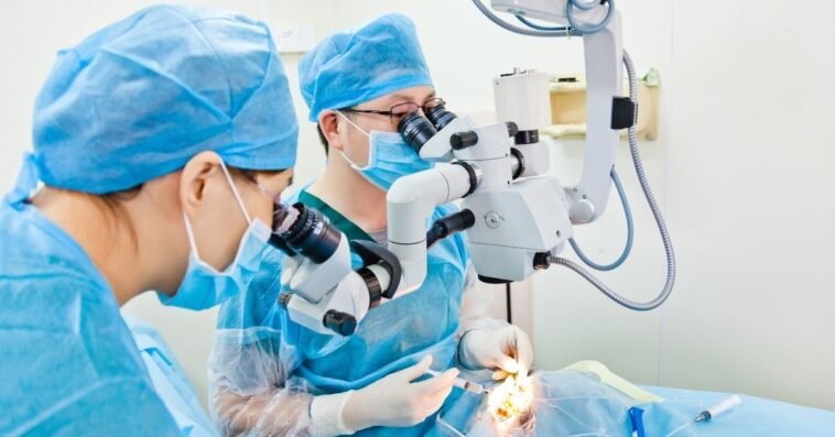
Eye surgery has advanced significantly over the past two decades. Today, most procedures, including cataract surgery, glaucoma treatment, and vision correction, are performed quickly and with minimal discomfort. In many cases, patients can return home the same day. These modern techniques help millions of people preserve and improve their vision.
This guide explains the most common types of eye surgery, how each one works, who may benefit, and what to expect before and after your procedure. Whether you are getting ready for surgery or just want to learn more about your options, you will find useful information to help you make informed decisions about your eye health.
1. Cataract Surgery
Cataract surgery is the most common eye operation performed worldwide. Cataracts form as a normal part of aging, causing the lens inside your eye to become cloudy. When the lens is cloudy, it cannot focus light properly, which can make your vision blurry or dim.
How Cataract Surgery Works
Cataract surgery involves three main steps. First, your eye doctor removes the cloudy lens using ultrasound or a laser. Then, a clear artificial lens, called an intraocular lens, or IOL is placed in your eye. This new lens remains in place and helps restore clear vision.
The procedure usually takes about 10 to 15 minutes. Your eye will be numbed to keep you comfortable, and you can return home the same day. Most people notice their vision improving within a day or two after surgery.
Types of Intraocular Lenses (IOLs)
Cataract surgery not only removes the cloudy lens, but it can also correct other vision problems, such as nearsightedness or astigmatism.
Monofocal IOLs (standard lenses)
Monofocal lenses are designed to focus at a single distance, most often for seeing distant objects clearly.
If you have a monofocal lens, you will likely still need reading glasses for close-up tasks, such as reading or sewing.
Toric IOLs
Toric lenses are designed for people with astigmatism, a condition in which the front of the eye (the cornea) is not perfectly round. These lenses help correct the uneven shape so you can see more clearly.
Multifocal and Trifocal IOLs
Multifocal and trifocal lenses are made to give you clear vision at near, middle, and far distances. With these lenses, you may not need glasses as often for reading or doing things up close.
Accommodating IOLs
Accommodating lenses help your eyes focus on both near and distant objects, just as your natural lens does.
Symptoms That Indicate You Need Cataract Surgery
You may need cataract surgery if you notice any of the following symptoms:
- Cloudy, dim, or blurry vision
- Poor night driving due to glare
- Halos around lights
- Difficulty reading or doing close work
- Sensitivity to bright light
Cataract surgery is usually recommended when your vision problems start to make daily activities difficult. Many people think they need to wait until the cataract is ‘ripe’ before having surgery, but this is not true.
2. YAG Laser Capsulotomy
Sometimes, months or even years after cataract surgery, your vision may become cloudy again. This is called posterior capsular opacification (PCO), or ‘secondary cataract.’ It is not the original cataract returning.
How YAG Laser Capsulotomy Works
Your eye doctor can treat this with a quick, painless laser procedure performed right in the office. The laser creates a small opening in the cloudy layer behind your lens implant, helping restore clear vision. Here is what you can expect:
- No incisions
- No downtime
- Immediate improvement in vision
Posterior capsular opacification happens in about one out of every four people after cataract surgery, but it can be easily treated with a short laser procedure.
3. Glaucoma Surgery
Glaucoma is a condition that can harm your optic nerve, often due to increased pressure inside the eye. If eye drops or laser treatments do not lower the pressure enough, your doctor may suggest surgery to help protect your vision.
Types of Glaucoma Surgery
1. SLT (Selective Laser Trabeculoplasty)
This procedure helps the eye drain fluid better in people with open-angle glaucoma.
SLT can lower the need for eye drops, and in some cases, you may be able to stop using them completely.
2. ALT (Argon Laser Trabeculoplasty)
ALT is an older type of laser treatment than SLT, but it is still used in some cases.
3. Laser Peripheral Iridotomy (LPI)
Laser peripheral iridotomy is done for people who have narrow angles in the eye, which raises the risk of a certain type of glaucoma.
During this procedure, the laser makes a tiny hole in the colored part of your eye (the iris) to help prevent a sudden rise in eye pressure.
B. Incisional Glaucoma Surgeries
1. Trabeculectomy
This surgery creates a small channel in the eye to help drain fluid and lower eye pressure.
Trabeculectomy is the standard surgery for people with advanced glaucoma.
2. Glaucoma Drainage Implants (Tube Shunts)
These are small silicone tubes that move fluid from inside the eye into a small reservoir, which helps lower eye pressure.
Glaucoma drainage implants are often used if trabeculectomy is not a good option or has not worked.
C. MIGS (Minimally Invasive Glaucoma Surgery)
MIGS stands for minimally invasive glaucoma surgery. These procedures have lower risks and usually allow for a quicker recovery than traditional glaucoma surgeries.
- iStent
- Hydrus Microstent
- Kahook Dual Blade
- OMNI system
MIGS procedures are often a good choice for people with mild to moderate glaucoma. In many cases, these surgeries can be done at the same time as cataract surgery.
4. Refractive Surgery
Refractive surgery changes the shape of your cornea or places a lens inside your eye to correct vision problems, such as:
- Nearsightedness (myopia)
- Farsightedness (hyperopia)
- Astigmatism
- Presbyopia
Most Common Types of Refractive Surgeries
LASIK (Laser-Assisted In Situ Keratomileusis)
LASIK is the most common type of refractive surgery worldwide.
During LASIK, your doctor creates a thin flap in the cornea, uses a laser to reshape the underlying tissue, and then replaces the flap.
PRK (Photorefractive Keratectomy)
Unlike LASIK, PRK does not involve creating a corneal flap.
Instead, the corneal surface is gently removed, and a laser reshapes the underlying tissue.
SMILE (Small Incision Lenticule Extraction)
In this procedure, a special laser creates a small disc, called a lenticule, inside the cornea. The lenticule is then removed through a tiny opening.
SMILE is designed to help most people recover quickly.
ICL (Implantable Collamer Lenses)
With ICL, your doctor places a soft, permanent lens inside your eye, while your natural lens stays in place.
ICL may be a good choice if you are very nearsighted or if you have dry eyes and cannot have LASIK.
5. Corneal Transplant Surgery (Keratoplasty)
During a corneal transplant, your doctor removes your damaged or cloudy cornea and replaces it with a healthy one from a donor.
This surgery can help restore vision in several conditions, including:
- Keratoconus
- Corneal scarring
- Corneal edema (Fuchs’ dystrophy)
- Severe infections
- Trauma
Types of Corneal Transplants
Penetrating Keratoplasty (PK)
This is a full-thickness corneal transplant, meaning the entire cornea is replaced.
DMEK (Descemet Membrane Endothelial Keratoplasty)
This procedure transplants only the innermost layer of the cornea.
DMEK often provides the sharpest vision and the fastest recovery among all types of corneal transplants.
DSAEK
DSAEK is a partial-thickness corneal transplant that replaces a slightly thicker layer than DMEK.
Corneal transplants are successful for most people, but regular follow-up visits are important because there is a small risk that your body could reject the new cornea.
6. Retina Surgeries
Retina surgery can help protect your vision if you have certain conditions, such as:
- Retinal detachment
- Diabetic retinopathy
- Macular hole
- Epiretinal membrane
- Retinal tears
Common Retina Procedures
Laser Photocoagulation
Laser treatment can seal leaking blood vessels or fix small tears in the retina.
Vitrectomy
A vitrectomy involves removing the gel-like substance inside your eye, called the vitreous, to treat conditions such as:
- Retinal detachment
- Macular holes
- Scar tissue
- Vitreous hemorrhage
Scleral Buckle
During a scleral buckle procedure, your doctor places a small silicone band around your eye. This band helps hold the retina in place as it heals.
Pneumatic Retinopexy
In pneumatic retinopexy, your doctor injects a small gas bubble into your eye. The bubble gently presses the retina back into position so it can heal.
7. Eye Muscle Surgery (Strabismus Surgery)
Eye muscle surgery is performed when the eyes are misaligned, a condition called strabismus.
The surgeon:
- Strengthens a muscle (resection)
- Weakens a muscle (recession)
This procedure helps:
- Correct eye alignment
- Reduce or eliminate double vision.
- Improve depth perception
This type of surgery can be performed in both children and adults.
8. Eyelid Surgery (Blepharoplasty)
Eyelid surgery is not just for cosmetic reasons. Sometimes, it is needed to improve vision or treat certain medical problems.
Blepharoplasty is performed to:
- Remove excess skin obstructing vision.
- Repair droopy eyelids (ptosis)
- Improve peripheral vision
- Treat eyelid tumors
Recovery after eyelid surgery is usually quick, and most people can get back to their normal activities soon after the procedure.
When Should You See an Eye Surgeon?
You should contact your eye doctor right away if you notice any of the following signs or symptoms:
- Sudden loss of vision
- Flashes or floaters
- Eye pain or pressure
- Distorted central vision
- Your vision has become cloudy, and it is hard to do your usual daily activities.
- Persistent double vision
- Your eyelids are drooping and blocking your vision. Seeking early treatment can help prevent permanent vision problems.
Conclusion
Eye surgery today is safer, faster, and more effective than ever before. Whether you have cataracts, glaucoma, vision changes, or retinal disease, modern treatments can help improve your vision and protect your overall eye health.
If you think you may need eye surgery or want a second opinion, schedule an appointment with your eye doctor. Getting checked early and starting treatment can lead to better results.
FAQ
1. What is the most common type of eye surgery?
Cataract surgery is the most common eye surgery worldwide. It replaces the cloudy natural lens with a clear artificial lens (IOL) and is considered one of the safest and most successful procedures in medicine.
2. How do I know if I need eye surgery?
Surgery may be necessary if you experience blurry vision affecting daily activities, persistent high eye pressure, sudden flashes or floaters, or loss of peripheral vision. An ophthalmologist will confirm the need based on a comprehensive eye exam.
3. Is eye surgery painful?
Most modern eye surgeries are not painful. Numbing drops or local anesthesia prevent discomfort, and patients typically describe the sensation as pressure rather than pain.
4. How long does recovery take after eye surgery?
Recovery varies by surgery:
- Cataract: 24–72 hours for noticeable improvement
- LASIK: Same day to 24 hours
- Corneal transplant: Several months
- Glaucoma surgery: A few weeks
- Retina surgery: Several weeks to months
Your doctor will give personalized instructions.
5. Are there risks with eye surgery?
All surgeries carry some risk, but severe complications are rare. Potential risks include infection, inflammation, pressure changes, retinal detachment, glare, or vision fluctuations. Selecting a qualified ophthalmologist significantly reduces these risks.
6. Can both eyes be operated on the same day?
For most procedures, only one eye is treated at a time. Some laser and refractive surgeries, such as LASIK, can be safely performed on both eyes during the same session.
7. Will I still need glasses after eye surgery?
The need for glasses depends on the type of surgery and the lens selection. Premium lenses (multifocal, toric, accommodating) can significantly reduce dependence on glasses, while standard lenses may still require glasses for reading.
8. What surgeries help avoid glasses?
LASIK, PRK, SMILE, ICL, and multifocal lens implants are the most effective procedures for reducing dependence on glasses.
9. Is cataract surgery permanent?
Yes. The cataract does not return. However, some individuals may develop posterior capsular opacification (PCO), which is easily treated with a YAG laser procedure.
10. What happens if I delay eye surgery?
Delaying surgery can:
- Make the surgery more difficult.
- Increase risks (especially in glaucoma or retinal detachment)
- Reduce the final visual outcome.
Consult your ophthalmologist if symptoms worsen.